Medical education faces a continuous challenge: providing students with highly detailed, realistic, and repeatable lab experiences while managing limited resources. For decades, physical cadaver laboratories have been the standard. However, specimen scarcity, the high costs of preservation, and health concerns regarding formaldehyde exposure are pushing institutions to seek better alternatives.
Today, top-tier medical universities are adopting digital solutions to bridge the gap between theoretical textbooks and practical, hands-on learning. Advanced visualization technologies are transforming how future healthcare professionals understand the complex networks of the human body.
Why Foundational Human Biology Matters
Before medical students can diagnose a patient or plan a surgical intervention, they must possess a deep comprehension of how the body operates. This requires studying the human body not just region by region, but as interconnected functional networks.
Systemic anatomy is a specialized branch of anatomical science that focuses on the study of the various systems within the human body. These networks, such as the skeletal, muscular, cardiovascular, and nervous systems, are examined in relation to their structure and function.
Unlike regional study, which emphasizes specific body areas and their localized components, systemic anatomy provides a holistic understanding of how different systems interact and operate to maintain the overall functionality of the body.
For medical students, grasping these concepts is non-negotiable. Understanding how the respiratory network supplies oxygen that the cardiovascular network then pumps to the muscular network is critical for clinical application. Without a firm grasp of systemic anatomy, diagnosing complex diseases becomes nearly impossible. It offers students and healthcare professionals a comprehensive framework for understanding the intricacies of bodily functions.
However, teaching these interconnected networks using traditional cadavers presents unique difficulties. Once a physical specimen is dissected to reveal the nervous network, it is incredibly difficult to reconstruct the surrounding muscular or vascular layers for a different lesson. This limitation hinders the student’s ability to repeatedly visualize how different networks overlap and interact in real-time.
The Shift Toward Digital Dissection Solutions
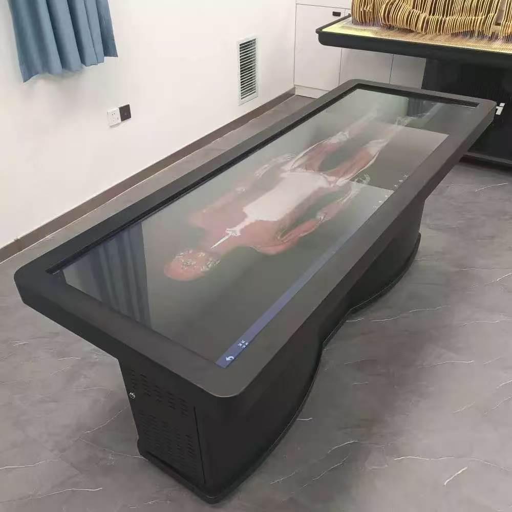
To overcome the physical limitations of traditional specimens, medical programs are increasingly integrating digital technology into their curriculum. A virtual dissection table replaces physical cadavers with highly accurate 3D simulations. This hardware and software integration allows educators to present complex biological structures in a controlled learning environment.
These digital platforms provide flexible access to anatomical models. Students can perform touch-based operations to rotate, zoom, and dissect structures layer by layer. If a student makes an error during a virtual dissection, they can simply reset the model and try again. This repeatable practice fosters deeper retention of complex spatial relationships without the fear of degrading a scarce physical specimen.
Furthermore, digital tables eliminate the biohazard risks and chemical exposure associated with traditional labs. Institutions save significantly on the long-term expenses related to cadaver procurement, specialized ventilation storage, and eventual disposal.
Elevating Instruction with the DIGIHUMAN Platform
For institutions seeking a high-quality digital transition, Institutions teaching systemic anatomy require tools that provide detailed anatomical visualization. DIGIHUMAN manufactures a Virtual Dissection Table designed specifically to meet the rigorous demands of medical universities and healthcare training facilities worldwide.
The platform is built upon real human tomographic sequence image data, entirely free of organic diseases or defects. It features ultra-high-precision anatomical data, utilizing over 17,571 cross-sections for male models and 16,141 for female models. Presented at a 0.1mm layer thickness, this level of detail allows learners to observe the most minute structures, from major organs down to tiny nerve endings and capillaries.
The software encompasses nine distinct body systems and displays the three-dimensional morphology of more than 6,000 anatomical structures. Instructors can utilize human peel and see-through functions to reveal structures from superficial skin down to deep organ layers. This specific capability makes teaching systemic anatomy significantly more intuitive, as students can easily trace a single nerve pathway or blood vessel through the entire virtual body.
Beyond 3D models, the platform integrates more than 1,700 CT and MRI images. These radiological scans correspond directly with the tomographic specimen images, bridging the gap between basic morphological learning and clinical diagnostic skills.
Interactive Features for Modern Classrooms
The user interface is designed for seamless classroom integration. Instructors can manipulate the life-sized models using touch controls, switching between backgrounds, adding custom 3D annotations, or utilizing transparency and stripping tools. Every major anatomical structure is marked with detailed annotations and corresponding textual interpretations, available in both Chinese and English.
The DIGIHUMAN platform also includes a wealth of supplementary resources. It features real dissection videos and 3D animations that visualize actual clinical procedures. For assessment, it provides a large repository of test questions and digital exercises to enhance knowledge consolidation.
The hardware is adaptable to various teaching environments. The touch screen can be lifted and tilted, accommodating different sightlines whether used in a large lecture hall or a small group skills lab. It also supports AR device compatibility, offering an even more immersive sensory experience for students.
By replacing limited physical resources with accurate, repeatable, and safe digital data, institutions can significantly enhance their curriculum. Adopting advanced digital platforms ensures that the next generation of physicians and researchers possess the deep structural knowledge required for superior patient care.
